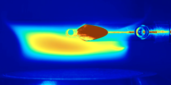
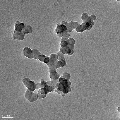
A widely employed technique for an imaging investigation of nanoparticles is electron microscopy. It is often used as a reference method for the verification of results obtained e.g. from optical measurement techniques. For microscopic investigations the particles first have to be sampled on appropriate sample holders. Especially particle sampling from high-temperature processes, e.g. like flame synthesis of nanoparticles, is challenging. Thermophoretic sampling of particles, which is based on particle movement caused by an external temperature gradient, is a common method to deposit particles on e.g. carbon based grids.
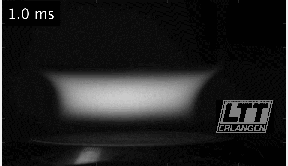
For representative results, however, the synthesis process must not be influenced significantly be the sampling process. Furthermore, sampling has to performed very quickly and reproducible due to the high temperatures and thus high thermal load on the sampling grids.
We investigated various propulsion technologies at the institute –mechanical, pneumatic and electromagnetic drives. Latest work reveals distinct advantages of electromagnetic drives; it allows for variable profiles of movement at high speeds with high temporal precision and accuracy. This enables tunable particle loadings of the grids for electron microscopy with a minimal influence of the particle synthesis process.
Another challenge is the analysis of the electron microscope images. In order to make statements about the size distributions of the aggregates and primary particles, a large number of images must be evaluated for example. For this purpose, new methods for semi-automatic image evaluation are developed at the institute including techniques such as image segmentation .
Literatur
- , , , :
Novel electric thermophoretic sampling device with highly repeatable characteristics
In: Review of Scientific Instruments 87 (2016), Article No.: 125108
ISSN: 1089-7623
DOI: 10.1063/1.4971988 - , , , , :
An optimized evaluation strategy for a comprehensive morphological soot nanoparticle aggregate characterization by electron microscopy
In: Journal of Aerosol Science 139 (2020), Article No.: 105470
ISSN: 0021-8502
DOI: 10.1016/j.jaerosci.2019.105470